WHAT IS GLAUCOMA?

Glaucoma is the name given to a group of eye disorders which are characterized by a progressive damage to optic nerve. Generally, there are no symptoms or warning signs of this disease and the patient himself does not get to know about the disease unless told by the doctor. This is because the sight loss is very gradual and starts in the periphery.

The primary problem in glaucoma is damage to the optic nerve. Intraocular pressure is the fluid pressure within the eye. The eye pressure at which optic nerve damage takes place is different for different individuals. Some individuals with high pressure do not develop nerve damage where as some with normal pressure can also develop optic nerve damage.

It is important to remember that while glaucoma is more common in older age group, it can occur at any age. Unfortunately there is currently no cure for Glaucoma and the vision lost due to it cannot be regained, although early detection and strict adherence to medication and halt or remarkably slow its progression.

UNDERSTANDING GLAUCOMA

The eye works very much like an old styled camera. The light comes in through the shutter, is focused by the lens, falls on the film and then we take it to be processed.

UNDERSTANDING GLAUCOMA

similarly, in the eye the light comes in through the cornea and pupil, focused by the lens onto the light sensitive layer i.e. Retina, and then goes via the Optic Nerve (Nerve of Sight) to the processor i.e. the Brain for processing. In individuals with Glaucoma, not all the image focused on Retina will be transferred to the Brain and hence result in progressive vision loss.

Although damage to the optic nerve is can be caused by decreased blood flow or injury, but the most common cause is increased intraocular pressure (IOP). IOP is generally controlled by circulation of aqueous, a fluid which bathes and nourishes the eye, keeping it firm and maintaining optimal eye pressure. Aqueous is produced by ciliary body, flows around the iris and drains into the trabecular meshwork. If there is disturbance in any of the above steps then it can result in a rise in IOP.

Glaucoma treatments in the form of eye drops, laser treatments or conventional surgery aim to reduce the IOP of eye.

WHO IS AT RISK OF GLAUCOMA?

Majority of patients with glaucoma will not experience any symptoms in the early stages of disease. Glaucoma is known as the ‘Silent thief of Sight’ as it starts affecting the peripheral vision before involving central vision. This lost peripheral vision is compensated by the other eye and hence the patient does not notice any defect.

The only way to know if you have glaucoma is by visiting an Ophthalmologist for a comprehensive eye examination.

Although anyone can have Glaucoma but there are few who are at a higher risk than others, these are those with :-

  • A family history of Glaucoma
  • High Eye Pressure
  • Age over 50 years
  • African or Asian ethnicity
  • Diabetes
  • Myopia
  • Previous History of Eye Injury
  • High or Low Blood Pressure

Most people are recommended to have a comprehensive eye examination for glaucoma every 2 years from age 50 or when advised by your eye health professional.

Patients of Asian ethnicity are recommended to have eye examination for Glaucoma starting at 40 years.

WHAT DOES TESTING OF GLAUCOMA ENTAIL?

The following tests will be completed by your ophthalmologist to assess ypour risk for Glaucoma.

  • IOP measurement. In Glaucoma the pressure within the eye is generally higher than normal. Glaucoma treatment currently focusses on lowering the IOP.
  • Assessment and Photography of Optic Nerve Head. Optic Nerve Head is the location where Optic Nerve enters the eye which in turn sends visual information to the Brain. In Glaucoma , Optic Nerve Head begins to demonstrate significant excavation as individual nerve fibres within it begin to die.
  • Assessment of Retinal Nerve Fibre Layer. There are a number of ways to measure the health of Nerve Fibre Layers. Modern technology utilizes Optical Coherence Tomography which can non invasively and accurately measure the thickness of nerve fibres. In Glaucoma, the RNFL thins out.
  • Assessment of your peripheral vision (Visual Field Testing). Visual fields are a formal measurement of the peripheral vision. In this test the patient is required to look straight ahead at the red light and press button when they see lights in their peripheral field. Visual field testing is very subjective does have a learning curve.
  • Assessment of Anterior Chamber Angle. The Anterior Chamber angle is the representation of drainage pathway of aqueous from the eye. Patients who have a narrow anterior chamber angle are more prone to have a high intraocular pressure. Its assessment is done by Gonioscopy.

Some rarer forms of Glaucoma such as Angle Closure Glaucoma can present with severe eye pain with redness and a decrease in vision. Such an eye condition is an emergency and needs urgent attention by ophthalmologist to lower the Intraocular Pressure and prevent permanent vision loss.

TYPES OF GLAUCOMA
GLAUCOMA SUSPECT

Clinicians refer to a patient as Glaucoma Suspect when they are showing signs of early Glaucoma but they are not yet sure. These signs can be a raised intraocular pressure but normal looking optic disc or it could be a patient with normal IOP but a suspicious looking optic disc. These individuals usually need to be monitored at regular intervals and no treatment is required till the time they are labelled as a Glaucoma suspect.

1.PRIMARY GLAUCOMAS

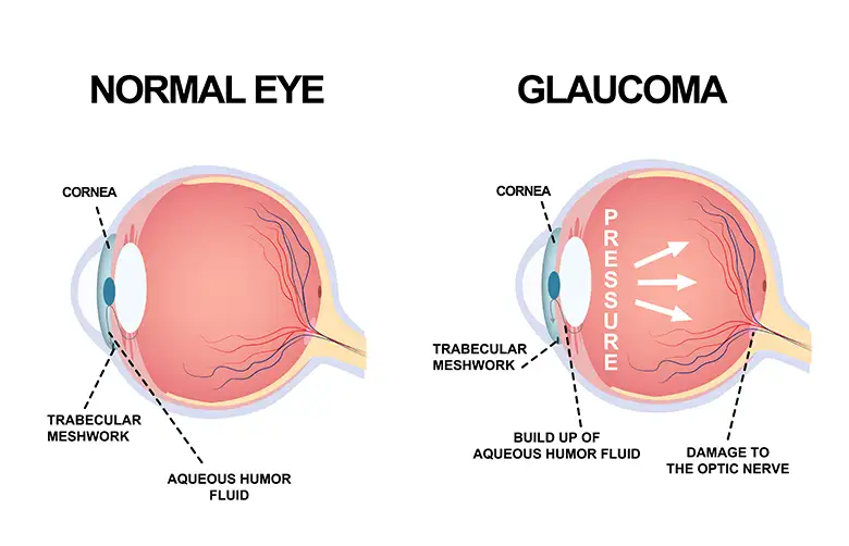
Primary open Angle Glaucoma is the most common type of Glaucoma in our population. In these cases the Optic Nerve is damaged usually due to a raised IOP. Most patients have no symptoms in the early stages as there is no pain and vision seems normal.

Normal Tension or Low Tension Glaucoma. In these individuals the IOP is normal but inspite of this they have damaged Optic Nerve. Evidence has shown that lowering of eye pressure further using eye drops or laser has stopped progression of Glaucoma in these patients.

Chronic Angle Closure Glaucoma. This is caused due to inherited narrowed down drainage pathways within the eye. This angle is located between the Iris and Trabecular meshwork. They can be helped by medicines and laser Treatment. Common amongst Asian and Chinese population.

Acute Angle Closure Glaucoma. This variant is a medical emergency which occurs due to obstruction of drainage pathway by the Iris. Patient presents with severe pain, redness, decreased vision , nausea and vomiting. Immediate treatment is warranted to prevent permanent damage.

Childhood Glaucoma. This is a rare form of Glaucoma caused due to abnormal drainage pathways. It can exist at birth or develop later. Parents may notice that the child has enlarged eyes, sensitive to light and has excessive watering.

2. SECONDARY GLAUCOMA

Glaucoma that develop due to other associated conditions of the eyes such as trauma, infection s, inflammations etc.

Various types are:

  • Traumatic Glaucoma
  • Steriod induced Glaucoma
  • Pseudoexfoliation Glaucoma
  • Pigmentary Glaucoma
  • Uveitis Glaucoma
  • Neovascular Glaucoma
  • Iridocorneal Endothelial Syndrome
GLAUCOMA IN FAMILIES

In many cases Glaucoma is an inherited (genetic) disease and is passed on from close relatives. Individuals with a direct relative ( parent, children, sibling) with Glaucoma have a much higher risk of developing Glaucoma – about 1 in 4 of such high risk cases go on to develop Glaucoma in their lifetime.

Recent TARRGET study has shown that the risk of developing Glaucoma in a family member increases once the affected member reaches severe stage of Glaucoma.

Thus it is very important for all patients diagnosed with glaucoma to alert first-degree relatives, and have the conversation to prompt them to have regular comprehensive eye examinations to screen for glaucoma.

I HAVE GLAUCOMA, WHAT NOW?

One of the most important contributions you can make once you have glaucoma is put your medications regularly as prescribed and attending scheduled review appointments. As Glaucoma treatment can get ineffective over time, ongoing testing is crucial to best protect your vision.

If you or a loved one has been diagnosed with Glaucoma you might be feeling overwhelmed and concerned for the future. Sardana Eye Institute is here to provide you with information you need so that you can learn how to manage the disease and take back control.

If you have been diagnosed with Glaucoma, you must periodically get your intraocular pressure, eye health and visual field checked.

DETECTION OF GLAUCOMA

Glaucoma is a complex disease and no single test can provide enough information to make a diagnosis. A regular eye checkup involves screening for Glaucoma and may indicate that further examinations are required. It is important to note that glaucoma blindness is irreversible. Therefore, early detection is crucial as glaucoma treatment can save remaining vision but it does not improve eyesight.

On referral to an eye specialist five tests are commonly done as a part of Glaucoma screening so as to formulate a management plan. The following is a brief overview of these 5 essential tests :

TONOMETRY measures the Intraocular Pressure. This is the only treatable aspect of Glaucoma management. Goldmann Applanation Tonometre is the most sensitive test to measure eye pressure.

OPHTHALMOSCOPY involves dilating the pupil and looking at the Optic Nerves. Any early damage can be suspected just looking at the Optic Nerves. It is important to document the cup:disc ratio.

PERIMETRY is done using automated field analysers. One eye is tested at a time to look for any peripheral visual field defects. These defects are the first to appear due to glaucoma damage which can only be documented with these tests and the patient himself does not notice it.

GONIOSCOPY is a test for evaluating the drainage angles of the eye. This test is essential to differentiate between Open and Closed Angle Glaucomas because their management is slightly different.

PACHYMETRY is used to measure the central corneal thickness. Any corneal thickness other than normal can influence tonometry readings.

TREATMENT OF GLAUCOMA

Although there is no cure for Glaucoma with treatment Glaucoma can be controlled in order to prevent any further sight loss.

Most individuals are able to manage their condition by using regular eye drops or some laser treatment. Information from tests, assessments and lifestyle, along with glaucoma type provides the information which will determine initial treatment.

Medications:

Medications help to either reduce the production of eye fluid or increase the drainage of this fluid or even both. The critical factor with medications is that it requires co-operation from patients.

Laser:

Laser surgery may be performed in certain conditions. Different lasers are used in Open and Closed angle Glaucomas. Lasers help in better flow of aqueous within the eye and augments its drainage.

Surgery:

Surgery may sometimes be required if the disease does not get controlled using above mentioned methods. Surgery creates an alternate pathway for the fluid to drain from the eye.

OUR TESTIMONIALS
OUR
VIDEOS

REQUEST AN APPOINTMENT

LET'S CONNECT NOW

Schedule your no obligation consultation by fill out the form below.